Artroscopia y ortopedia

Artroscopia y ortopedia
Dr Benjamín Pineda

viernes, 22 de noviembre de 2013

Viscosupplementation for Knee Osteoarthritis

Viscosupplementation is a medical procedure during which lubricating fluid is injected into a joint. Also called hyaluronic acid injections or hyaluronan injections, viscosupplementation is most commonly used to treat symptoms of knee osteoarthritis.
Hyaluronic acid is a key component of the joint fluid in healthy joints, but is found in lower concentrations in osteoarthritic joints
By adding hyaluronic acid to the existing joint fluid of an osteoarthritic knee, the goal is to:
  • Facilitate better knee movement
  • Reduce pain
  • Perhaps slow osteoarthritis progression
Typical candidates for viscosupplementation are people with knee osteoarthritis who have failed to improve with other non-surgical treatments.
Following the injections, it is generally recommended that patients engage in a rehabilitation program that includes gentle, progressive knee exercise. The goals of rehabilitation are to improve range of motion and develop muscle strength to support the knee.

Knee exercises for Osteoarthritis of Knee

http://doctorsvideos.blogspot.mx/2011/05/knee-exercises-for-osteoarthritis-of.html
http://youtu.be/LmUJAqE4uLU


Basic isometric exercises for those who suffer from osteoarthritis for the quadriceps as well as Hip Abductors

Operado Ruiz de Galarreta, al que le quedan cinco meses de baja

http://www.canalathletic.com/noticias/2013-11-21/operado-ruiz-galarreta-quedan-201311211455.html


CEDIDO AL MIRANDÉS
El mediocentro del Athletic ha sido intervenido en Vitoria del ligamento cruzado anterior de su rodilla derecha casi un mes después de lesionarse
21 de noviembre de 2013


Operado Ruiz de Galarreta, al que le quedan cinco meses de baja
Ruiz de Galarreta abandona el campo lesionado tras el Mirandés y Numancia del 27 de octubre.
JAVIER ORTIZ DE LAZCANO | BILBAO.-
Casi un mes después de lesionarse el pasado 26 de octubre, Íñigo Ruiz de Galarreta (Eibar, 20 años) ha sido operado hoy por el doctor Mikel Sánchez en la Clínica USP de Vitoria del ligamento cruzado anterior de su rodilla derecha. La intervención, realizada mediante artroscopia, ha transcurrido sin complicaciones.
El tiempo transcurrido entre el percance y el quirófano se explica porque el traumatólogo considera necesario fortalecer los músculos de la zona antes de operar.
El mediocentro cedido por el Athletic al Mirandés se lesionó en el partido ante el Numancia. Es la segunda rodilla en la que sufre una grave lesión. La pasada campaña fue intervenido de la izquierda. Le quedan alrededor de cinco meses de baja.

miércoles, 20 de noviembre de 2013

¿ Que proponen ?

Masculino de 28 años fractura expuesta de tibia gustillo iiia po de reducción con clavo use coloca injerto autologo se cita acude 8. Semanas con pus cultivó e coli ya se lavó y actualmente limpio q proponen

domingo, 17 de noviembre de 2013

“El CS logra reducir casi un 25% el riesgo de prótesis en pacientes con artrosis de rodilla”

http://www.condroprotectores.es/el-condroitin-sulfato-logra-reducir-casi-un-25-el-riesgo-de-protesis-en-pacientes-con-artrosis-de-rodilla/


Dr. Arden: “El CS logra reducir casi un 25% el riesgo de prótesis en pacientes con artrosis de rodilla”

En España se estima que el 12% de las personas de más de 60 años sufre artrosis de rodilla y en muchos casos acaban desarrollando un deterioro de la articulación que hace que acaben necesitando una prótesis. Sin embargo, según se desprende de los resultados de un nuevo estudio de la Universidad de Oxford (Reino Unido) sobre la eficacia del condroitín sulfato, el uso de este fármaco permitiría reducir hasta casi un 25% el riesgo de recibir una prótesis.
Dr. Nigel Arden
Dr. Nigel Arden, reumatólogo Universidad de Oxford
Con motivo del Congreso Nacional de la Sociedad Española de Médicos de Atención Primaria (SEMERGEN), que se celebró la semana pasada en Barcelona, uno de sus autores, el prestigioso reumatólogo Nigel Arden, explicó a Europa Press la importancia de evitar que los pacientes con artrosis de rodilla reciban una prótesis, ya que “el 52% de quienes la reciben admiten que suele tener un gran impacto en sus vidas“. Además, apunta este experto, la realización de una artroplastia total de rodilla también tiene un importante impacto económico en el sistema sanitario.
Ante este doble impacto, en el estudio presentado Arden y su equipo pretendían ver si el uso del condroitín sulfato podía reducir el riesgo de prótesis en pacientes con artrosis de rodilla ya diagnosticada. Para ello, recurrieron a la base de datos del Sistema de Información para el Desarrollo de la Investigación en Atención Primaria (SIDIAP) de Cataluña, que incluye información de Atención Primaria y facturación de farmacia.
En total, participaron en el estudio 122.633 pacientes, de los que el 6,8% acabaron necesitando una prótesis de rodilla. Entre los pacientes que fueron tratados con condroitín sulfato, se observó como el riesgo de necesitar una prótesis se redujo a los seis meses de iniciar el tratamiento, según asegura Arden. En concreto, el uso de este tratamiento disminuyó un 13% el riesgo de prótesis, un beneficio “importante” que además se incrementó a medida que se prolongaba el uso del tratamiento. De hecho, tras al menos un año de tratamiento, el riesgo de prótesis se redujo en un 23%. “Los resultados son importantes, sobre todo si se tiene en cuenta que las osteoartritis son los tipos más frecuentes de artritis“.

jueves, 14 de noviembre de 2013

Pulmonary Embolism After Total Knee Arthroplasty

http://icjr.net/report_100_east_posters.3.htm#.UoUcahpyHsa


Reports ICJR Meetings

Award-Winning Posters from ICJR East

Five posters were recognized with ICJR East Abstract Awards.
By ICJR Staff - October 14, 2013
Pulmonary Embolism After Total Knee Arthroplasty

Primary Author: Nicholas B. Schraut

Institution: University of Illinois at Chicago – Department of Orthopaedic Surgery, Chicago, IL, USA

Co-Authors: Vincent Moretti (University of Illinois at Chicago – Department of Orthopaedic Surgery, Chicago, IL, USA), Ritesh Shah (Illinois Bone and Joint Institute, Morton Grove, IL, USA)
INTRODUCTION: Pulmonary embolism (PE) is a rare but potentially devastating complication of total knee arthroplasty (TKA). The purpose of this study was to assess recent national trends in PE occurrence after TKA and evaluate patient outcomes.
METHODS: International Classification of Disease-9th Revision (ICD-9) procedure codes were used to search the National Hospital Discharge Survey (NHDS) for patients admitted to U.S. hospitals after primary TKA for the years 2001-2010. ICD-9 diagnosis codes were used to identify patients who developed an acute PE during the same admission.
Data regarding patient demographics, hospitalization length, discharge disposition, lower-extremity deep vein thrombosis (DVT), mortality, and hospital size/location were gathered. Trends were evaluated by linear regression with Pearson’s correlation coefficient(r), and statistical comparisons were made using Student’s t-test, z-test for proportions, and chisquare analysis with a significance level of 0.05.
RESULTS: 35,220 patients admitted for primary TKA were identified; 159 (0.045%) of these patients developed an acute PE during the same admission. After adjusting for fluctuations in annual TKA performed, the development of PE after TKA demonstrated a weak negative correlation with time (r=0.17, Figure 1), insignificantly decreasing from an average rate of 0.049% between 2001 and 2005 to 0.041% between 2006 and 2010 (p=0.26). The size of the hospital was found to significantly impact the incidence of PE and primary TKA, with the lowest rate seen in hospitals under 100 beds (0.23%) and the highest rate seen in those with over 500 beds (0.65%, p=0.01). No significant differences in PE incidence were noted based on U.S. region (p=0.38).
The mean age of patients with PE was 67.7 years. This group included 54 men and 105 women. The non-PE group had a mean patient age that was insignificantly lower at 66.6 years (p=0.21) and included 12,450 men and 22,611 women. Gender was also not significantly different (p=0.68) between PE groups. The number of medical comorbidities was significantly higher in those with PE (mean 6.42 diagnoses) than those without PE (mean 4.89 diagnoses, p<0.01). Average hospitalization length also varied based on PE status, with significantly longer stays for those with PE (8.2 days versus 3.7 days, p<0.01). The rate of DVT was higher in the PE group (12.7% versus 0.48%, p<0.01). Mortality was also significantly higher for the PE group (3.9% versus 0.09%, p<0.01). Discharge disposition did not significantly vary based on PE status, with 61.5% of PE patients and 64.0% of non-PE patients able to go directly home (p=0.59) after their inpatient stay.
DISCUSSION/CONCLUSION: This study demonstrates that PE can have a significant impact on patient outcomes and healthcare costs, with associated 43-fold increase in mortality and a doubling of the inpatient admission duration. Additionally, although the risk of PE after primary TKA remains rare, efforts to prevent or minimize this complication over the last 10 years have not had a significant impact on its occurrence. This risk of PE appears to be greatest in patients with multiple medical comorbidities and established DVTs. Interestingly, the PE rate also demonstrated variability based on hospital size.

miércoles, 13 de noviembre de 2013

XXV Congreso Mexicano de Ortopedia y Traumatología, México, 2014


XXV Congreso Mexicano de Ortopedia y Traumatología, México, 2014


Los pacientes portadores de prótesis totales de cadera metálicas no tienen un mayor riesgo de cáncer que los portadores de prótesis con superficies de apoyo no metálicas

http://www.bmj.com/content/344/bmj.e2383


Los pacientes portadores de prótesis totales de cadera metálicas no tienen un mayor riesgo de cáncer que los portadores de prótesis con superficies de apoyo no metálicas.

BMJ, 03/04/2012, 

Risk of cancer in first seven years after metal-on-metal hip replacement compared with other bearings and general population: linkage study between the National Joint Registry of England and Wales and hospital episode statistics”.


La prótesis total de cadera se asocia con la liberación de cobalto, cromo y molibdeno y hay evidencias de que la exposición elevada a los iones metálicos se asocia con un aumento de la incidencia de ciertos cánceres. Este estudio británico pretende determinar si el uso de superficies de soporte de metal-metal se asocia con un aumento del diagnóstico de cáncer en los primeros años después del reemplazo total de cadera y, específicamente, con un aumento en el melanoma maligno, de próstata, hematológicos y de las vías urinarias. Se siguió a 40.576 pacientes con reemplazo de cadera de superficies de contacto metal-metal y 248.995 con rodamientos de distinto material. Se observó que la incidencia de nuevos diagnósticos de cáncer fue baja después de la artroplastia de cadera (1,25% en un año, 95% IC 1,21% a 1,30%) y menor que el previsto de la misma edad y sexo de la población normal (1,65%, 1,60% a 1,70%). Comparando con los rodamientos de materiales alternativos, no se encontró evidencia de que la superficie metal-metal se asociara con un mayor riesgo de cualquier diagnóstico de cáncer en los siete años después de la cirugía, con una media de seguimiento de tres años (67.361) de los pacientes observados durante cinco años o más. Tampoco hubo un aumento en el riesgo de melanoma maligno, cáncer de próstata, hematológico o cánceres de las vías urinarias. Estos datos son alentadores, pero son resultados de un estudio observacional con seguimiento a corto plazo. Así, es necesario seguir investigando los efectos de la exposición a metales ortopédicos a más largo plazo, puesto que algunos tipos de cáncer tienen un largo período de latencia.


Risk of cancer in first seven years after metal-on-metal hip replacement compared with other bearings and general population: linkage study between the National Joint Registry of England and Wales and hospital episode statistics

Abstract

Objective To determine whether use of metal-on-metal bearing surfaces is associated with an increased risk of a diagnosis of cancer in the early years after total hip replacement and specifically with an increase in malignant melanoma and haematological, prostate, and renal tract cancers.
Design Linkage study with multivariable competing risks flexible parametric survival model to examine the incidence of new diagnoses of cancer in patients with metal-on-metal hip replacement compared with those with alternative bearings and to compare the observed incidence of diagnoses in patients undergoing hip replacement with that predicted by national incidence rates in the general population.
Setting National Joint Registry of England and Wales (NJR) linked to NHS hospital episode statistics data.
Participants 40 576 patients with hip replacement with metal-on-metal bearing surfaces and 248 995 with alternative bearings.
Main outcome measures Incidence of all cancers and incidence of malignant melanoma and prostate, renal tract, and haematological cancers.
Results The incidence of new diagnoses of cancer was low after hip replacement (1.25% at one year, 95% confidence interval 1.21% to 1.30%) and lower than that predicted from the age and sex matched normal population (1.65%, 1.60% to 1.70%). Compared with alternative bearings, there was no evidence that metal-on-metal bearing surfaces were associated with an increased risk of any cancer diagnosis in the seven years after surgery (mean follow-up of three years, 23% (n=67 361) of patients observed for five years or more). Similarly, there was no increase in the risk of malignant melanoma or haematological, prostate, and renal tract cancers. The adjusted five year incidence of all cancers for men aged 60 was 4.8% (4.4% to 5.3%) with resurfacing, 6.2% (5.7% to 6.7%) with stemmed metal-on-metal, and 6.7% (6.5% to 7.0%) for other bearing surfaces. Equivalent rates for women aged 60 were lower: 3.1% (2.8% to 3.4%) with resurfacing, 4.0% (3.7% to 4.3%) with stemmed metal-on-metal, and 4.4% (4.2% to 4.5%) with other bearings.
Conclusions These data are reassuring, but the findings are observational with short follow-up. The use of hospital episode statistics data might underestimate cancer diagnoses, and there is the possibility of confounding by indication. Furthermore, as some cancers have a long latency period it is important that we study the longer term outcomes and continue to investigate the effects of exposure to orthopaedic metals

martes, 12 de noviembre de 2013

Metallosis after knee replacement

http://www.kneeguru.co.uk/KNEEnotes/primers/joint-cartilage-and-arthritis-primer/metallosis-after-knee-replacement

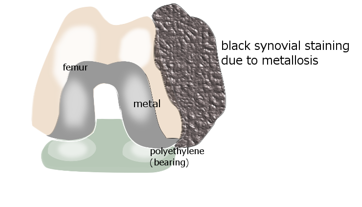
Synovial staining due to metallosis
© KNEEguru
Metallosis is a syndrome associated with metal joint implants, where friction between two metal surfaces releases metal ions (electrically charged particles) into the joint, and these trigger an inflammatory response. The term 'metal-induced synovitis' describes those parts of the synovial joint lining that bloat up and become rubbery and filled with a florid black staining due to the metal ions.
Within the knee this condition is usually related to a previous knee replacement (usually a total knee replacement, but also sometimes in a partial/unicompartmental1 knee replacement) in which the polyethylene components have worn down and inadvertently allowed metal components to come into contact where they normally would not. In particular it is often the polyethylene part of the metal-backed patella that is at fault, where the polyethylene breaks down and allows the back of the patellar component to make friction contact with the metal femoral component2. Metal-backed patellar components are no longer in frequent use but of course there are still many patients who had had this type of implant done in previous years. Mechanisms of failure of the polyethylene include polyethylene wear, fracture of the polyethylene or the underlying bone, and dissociation of the polyethylene from its metal backing.
The condition is more frequently recognised in hip replacements. Although rare in the knee, metallosis is a diagnosis must be thought of when investigating a patient whose knee is not settling after a knee replacement

Components of a knee replacement

Knee replacements are not all the same. Different manufacturers have their own designs, materials and components, and the surgeon will make his choice among their products based on his/her assessment of the patient's needs and his/her own clinical experience. However, most knee replacements contain -
  • a metal femoral component
  • a metal tibial component
  • a 'bearing' between them of high density polyethylene, which may be mobile or attached to the tibial component
  • a patellar component of polyethylene or metal-backed polyethylene
In uncemented components there may be coatings of various kinds at the back of the components to encourage bone adhesion.
The metals from which the implant is manufactured may include3 -
  • titanium and titanium alloys
  • cobalt and cobalt-chrome alloys. Cobalt chrome alloys contain a nickel component, which is very relevant in people who are allergic to nickel.
  • zirconium and zirconium alloys
  • ceramics including oxidised zirconium
  • tantalum
Hip implants may be metal-on-polyethylene, metal-on-metal, metal-on-ceramic or ceramic-on ceramic, but most knee implants are metal-on-polyethylene. The issue of metal-on-metal designs is current and controversial4. Several hip implant manufacturers have recalled their metal-on-metal implants5, and patients who have already received such implants are carefully monitored and may be subjected to additional surgery to replace them
In the knee, which is what we are talking about here, we are not talking about metal-on-metal designs but to inadvertent metal/metal contact due to failure of the polyethylene bearing between the metal components. The metal ions thereby released may cause both local and wider (systemic) symptoms.

Local and systemic effects of metal toxicity

There seems to be two types of problems related to the situation we are discussing. On the one hand the polyethylene wear leaves real particulate debris of polyethylene, which is taken up by the lymph cells and transported to the lymph glands. As the body tries to rid itself of these particles a process called 'osteolysis' may occur, where bone is broken down and weakened in areas around the implant. This can be seen on X-ray or CT imaging.
On the other hand, the breakdown of the polyethylene may distort the mechanics of the implant and allow metal to come into contact with metal. The resultant friction may release charged metal ions, which are not the same as the particulate matter of the polyethylene, although sometimes, but rarely, there may be actual metal particles. The presence of metal ions can be inferred from X-ray changes which we will discuss later.
The breakdown of the polyethylene may affect the mechanics of the implant, the osteolysis may allow loosening of the implant, the debris may bring other inflammatory-response cells into the area which will be releasing chemicals of their own. The metal ions are taken up by the synovium(the joint lining), which may become abnormal - bloated in patches and filled up with grey-black staining. Uptake by histiocytes1 may affect regional lymph glands, and metal ions in the bloodstream may cause metal toxicity.
The combination of all of these factors may lead to2 -
  • severe knee pain and quite marked knee swelling, with excessive fluid in the joint
  • crepitus, and feelings of instability associated with loosening of the implant
  • pseudotumours6
  • fractures around the implant
  • skin rashes, confusion, tinnitus, deafness, and even dementia.
Some patients may have a frank allergy to certain metals, eg nickel,cobalt and chrome while in other patients it may be more a matter of metal toxicity. Skin patch testing may differentiate between the metals, although it may be necessary to do blood tests and histology on the lymph nodes. Potentially the most harmful components are cobalt from cobalt-chromium alloy, nickel from stainless steel, and vanadium from titanium alloy7.
X-rays may show excessive fluid within the joint, osteolysis around the implant.  The joint lining andsuprapatellar pouch may show a fine linear radio-opacity called a 'metal-line. Other radiographic signs called 'clouds' and 'bubbles'2 may be indicative of the metal within the synovium as well as the florid synovial outgrowth, seen during surgery as areas of joint lining (synovium) heaped up into what look like soft black lumps.

Blood levels

It would seem logical to simply monitor knee replacement patients for raised metal levels, but this is not as simple as it seems. The biggest culprit for toxicity seems to be cobalt-chrome but there is no generally accepted threshold beyond which serum or blood concentrations of cobalt and chromium are known to be toxic8. Despite this, it appears that a revision from a cobalt-chrome femoralcomponent to a ceramic one, or replacement of a metal-backed, with a polyethylene patellar component should be contemplated when there appears to be toxicity problems9.
Ceramic components appear to offer advantages, but the long term outcome is still being evaluated10.

References

1. http://ispub.com/IJPA/4/2/10194

M den Bakker, R Brouwer. Metallosis In Revision Knee Arthroplasty. The Internet Journal of Pathology Volume 4 Number 2.
2. http://www.ncbi.nlm.nih.gov/pmc/articles/PMC3303397/

Romesburg JW, Wasserman PL, Schoppe CH. Metallosis and Metal-Induced Synovitis FollowingTotal Knee Arthroplasty: Review of Radiographic and CT Findings. ​J Radiol Case Rep. 2010;4(9):7-17. doi: 10.3941/jrcr.v4i9.423. Epub 2010 Sep 1.
5. http://www.kcrlegal.com/hip-replacement-metal-ions.aspx

DePuy ASR metal hip replacement devices linked to metal toxicity in the bloodstream.
6. http://hosp.gcnpublishing.com/fileadmin/qhi_archive/ArticlePDF/AJO/03702...

F. Daniel Kharrazi, MD, Benjamin T. Busfield MS, MD, Daniel S. Khorshad, Francis J. Hornicek MD, PhD, and Henry J. Mankin, MD. Knee Mass From Severe Metallosis After Failure of a Metal-Backed Patellar Component Total Knee Arthroplasty
7. http://www.bjj.boneandjoint.org.uk/content/63-B/3/435.long

Rae T. The toxicity of metals used in orthopaedic prostheses. An experimental study using cultured human synovial fibroblasts. J Bone Joint Surg Br. 1981;63-B(3):435-40.
8. http://www.youtube.com/watch?v=NGZV7ofdvj8

Metal Toxicology Expert Part 5: What studies have been conducted regarding cobalt exposure?
9. http://www.ncbi.nlm.nih.gov/pubmed/23590796

Thakur RR, Ast MP, McGraw M, Bostrom MP, Rodriguez JA, Parks ML. Severe persistent synovitis after cobalt-chromium total knee arthroplasty requiring revision. Orthopedics. 2013 Apr;36(4):e520-4. doi: 10.3928/01477447-20130327-34.
10. http://www.ncbi.nlm.nih.gov/pubmed/22582104

Bergschmidt P, Bader R, Ganzer D, Hauzeur C, Lohmann C, Rüther W, Tigani D, Rani N, Prats FL, Zorzi C, Madonna V, Rigotti S, Benazzo F, Rossi SM, Kundt G, Bloch HR, Mittelmeier W. Ceramic femoral components in total knee arthroplasty - two year follow-up results of an international prospective multi-centre study. Open Orthop J. 2012;6:172-8. doi: 10.2174/1874325001206010172. Epub 2012 Apr 20.
ABOUT THE AUTHOR

Dr Sheila Strover

Clinical Editor
Degrees: 
BSc (Hons)
MB BCh
MBA
Dr Sheila Strover is the founder of the KNEEguru website. Although not a knee surgeon, she has a sound understanding of knee surgery and...

domingo, 10 de noviembre de 2013

What to Do if Your Child Tears an A.C.L.


Artroscopia de rodilla

http://orthoinfo.aaos.org/topic.cfm?topic=A00619


Artroscopia de rodilla (Knee Arthroscopy)
La artroscopia es un procedimiento quirúrgico común en el cual una articulación (artro-) se visualiza (-scopia) usando una cámara pequeña. La artroscopia da a los médicos una visión clara del interior de la rodilla. Esto los ayuda a diagnosticar y tratar los problemas de rodilla.
Los avances técnicos han llevado a monitores de alta definición y a cámaras de alta resolución. Éstas y otras mejoras han transformado a la artroscopia en una herramienta muy eficaz para tratar los problemas de rodilla. Según la Sociedad Ortopédica Americana para la Medicina del Deporte, más de 4 millones de artroscopias de rodilla se realizan en todo el mundo cada año.
Descripción
La artroscopia se realiza a través de pequeñas incisiones. Durante el procedimiento, su cirujano ortopédico inserta el artroscopio (un instrumental con una cámara pequeña del tamaño de un lápiz) en la articulación de su rodilla. El artroscopio envía la imagen al monitor de una televisión. En el monitor, su cirujano puede ver las estructuras de la rodilla con gran detalle.
Su cirujano puede usar la artroscopia para sentir, reparar o quitar tejido lesionado. Para hacerlo, se insertan pequeños instrumentos quirúrgicos a través de otras incisiones alrededor de su rodilla.
Preparación para la cirugía

Figura artroscópica de ligamento cruzado anterior roto [estrella amarilla]
Si usted decide tener una artroscopia de rodilla, puede necesitar un examen físico completo con su médico de familia antes de la cirugía. Su médico evaluará su salud e identificará cualquier problema que pueda interferir con su cirugía.
Antes de la cirugía, informe a su cirujano ortopédico acerca de cualquier medicamento o suplemento que usted toma. El médico le dirá qué medicamentos usted debe dejar de tomar antes de la cirugía.
Para ayudar a planificar el procedimiento, su cirujano ortopédico puede pedir exámenes preoperatorios. Éstos pueden incluir conteos de células de la sangre o un EKG (electrocardiograma).
Cirugía
Casi toda la cirugía artroscópica de rodilla se realiza en atención ambulatoria.

Llegada

Su hospital o centro quirúrgico lo contactarán para darle los detalles específicos de su cita. Probablemente le pedirán que llegue al hospital una hora o dos horas antes de la cirugía. No coma ni beba en absoluto después de la medianoche antes de la cirugía.

Anestesia

Cuando usted llega para la cirugía, un miembro del equipo de anestesia hablará con usted. La artroscopia puede realizarse bajo anestesia local, regional o general.
  • La anestesia local adormece sólo su rodilla
  • La anestesia regional adormece su cuerpo por debajo de la cintura
  • La anestesia general lo hace dormir totalmente
El anestesista lo ayudará a decidir qué método sería mejor para usted.
Si le realiza anestesia local o regional, usted tal vez pueda ver el procedimiento en el monitor de una televisión.

Procedimiento

El cirujano ortopédico hará unas incisiones pequeñas en su rodilla. Una solución estéril será usada para llenar la articulación de la rodilla y quitar cualquier líquido turbio. Esto ayuda a que su cirujano ortopédico vea su rodilla con claridad y con todos los detalles.

Artroscopia de rodilla

Primer plano de reparación de un menisco
La primera tarea de su cirujano es diagnosticar debidamente su problema. Él o ella insertará el artroscopio y usará la imagen proyectada en la pantalla para guiarlo. Si el tratamiento quirúrgico es necesario, su cirujano insertará instrumentos diminutos a través de otra pequeña incisión. Estos instrumentos pueden ser tijeras, rasuradores o láseres.
Esta parte del procedimiento generalmente dura de 30 minutos a un poco más de una hora. El tiempo que dure depende de los hallazgos y del tratamiento necesario.
La artroscopia de rodilla se usa más comúnmente para:
  • Quitar o reparar cartílago de meniscos rotos
  • Reconstruir un ligamento cruzado anterior roto
  • Recortar pedazos rotos de cartílago articular
  • Quitar fragmentos sueltos de hueso o cartílago
  • Quitar tejido sinovial inflamado
Su cirujano puede cerrar las incisiones con una sutura o con steri-strips (bandas adhesivas pequeñas) y cubrirlas con un vendaje suave.
Usted será trasladado a la sala de recuperación y podría irse a su casa en un plazo de 1 a 2 horas. Asegúrese de que haya alguien con usted para que lo lleve a su casa.
Recuperación
La recuperación de la artroscopia de rodilla es mucho más rápida que la cirugía tradicional a rodilla abierta. De todos modos es importante seguir las instrucciones de su cirujano ortopédico cuidadosamente después que usted regresa a su casa. Usted debe pedir a alguien que lo acompañe la primera noche en su casa.

Inflamación

Mantenga su pierna elevada lo más posible durante los primeros días después de la cirugía. Aplique hielo conforme a las recomendaciones de su médico para aliviar la inflamación y el dolor.

Cuidado de las compresas

Usted dejará el hospital con un vendaje que cubrirá su rodilla. Mantenga las incisiones limpias y secas. Su cirujano le dirá en qué momento se podrá duchar o bañar, y cuándo debería cambiar el vendaje.
Su cirujano lo verá en el consultorio unos pocos días después de la cirugía para controlar su progreso, revisar los resultados de la cirugía y comenzar su programa de tratamiento postoperatorio.

Soportar peso

La mayoría de los pacientes necesitan muletas u otro tipo de asistencia después de la cirugía artroscópica. Su cirujano le dirá cuándo es seguro soportar peso en su pie y pierna. Si usted tiene alguna pregunta acerca de soportar peso, llame a su cirujano.

Conducir vehículos

Su médico discutirá con usted en qué momento usted podrá conducir. Esta decisión se basa en un número de factores, incluyendo:
  • La rodilla involucrada
  • Si usted conduce un vehículo de cambios automáticos o manuales
  • La naturaleza del procedimiento
  • Su nivel de dolor
  • Si usted está usando medicamentos narcóticos para el dolor
  • Cuánto usted puede controlar su rodilla.
Típicamente los pacientes pueden conducir entre 1 y 3 semanas después del procedimiento.

Medicamentos

Su médico le recetará un medicamento para el dolor para ayudar a aliviar la incomodidad después de la cirugía. Su médico también puede recomendarle medicamentos como aspirina para disminuir el riesgo de coágulos sanguíneos.

Ejercicios para fortalecer su rodilla


Usted debe ejercitar su rodilla regularmente durante varias semanas después de la cirugía. Esto restablecerá el movimiento y fortalecerá los músculos de su pierna y rodilla.
El ejercicio terapéutico jugará un rol importante en su recuperación. Un programa de terapia física formal puede mejorar el resultado final.
Complicaciones y signos de advertencia
Como ocurre con cualquier cirugía, hay riesgos asociados con la artroscopia de rodilla. Éstos ocurren de manera infrecuente y son menores y tratables.

Complicaciones

Los problemas postoperatorios potenciales con la artroscopia de rodilla incluyen:
  • Infección
  • Coágulos sanguíneos
  • Acumulación de sangre en la rodilla

Signos de advertencia

Llame a su cirujano ortopédico inmediatamente si usted experimenta cualquiera de los síntomas siguientes:
  • Fiebre
  • Escalofríos
  • Zona de calor o enrojecimiento persistente alrededor de la rodilla
  • Dolor persistente o en aumento
  • Inflamación significativa en su rodilla
  • Dolor en aumento en el músculo de su pantorrilla
Resultado final
A menos que a usted le realicen una reconstrucción de ligamentos, usted debería poder regresar a la mayoría de sus actividades físicas después de 6 a 8 semanas, o a veces mucho antes. Las actividades de más alto impacto deben evitarse durante un tiempo más largo. Usted necesitará hablar con su médico antes de retomar actividades físicas intensas.
Si su trabajo implica tareas pesadas, puede pasar más tiempo antes que usted pueda volver a su trabajo. Discuta con su médico cuándo es razonable y seguro que usted vuelva a trabajar.
El resultado final de su cirugía probablemente será determinado por el grado de daño en su rodilla. Por ejemplo, si el cartílago articular en su rodilla se ha desgastado completamente, entonces la recuperación completa puede que no sea posible. Usted podría necesitar cambiar su estilo de vida. Esto puede significar limitar sus actividades y buscar alternativas de ejercicios de bajo impacto.
Last reviewed: Marzo 2010
AAOS does not endorse any treatments, procedures, products, or physicians referenced herein. This information is provided as an educational service and is not intended to serve as medical advice. Anyone seeking specific orthopaedic advice or assistance should consult his or her orthopaedic surgeon, or locate one in your area through the AAOS "Find an Orthopaedist" program on this website.

Refijación de arrancamiento del ligamento cruzado posterior con fragmentos óseos tibiales por medio de una placa gancho

http://www.elsevierinstituciones.com/ei/ctl_servlet?_f=1012&id_articulo=90122527&idinst=219

  1. Técnicas Quirúrgicas en Ortopedia y Traumatología

    Vol. 21. Núm. 01. Enero - Marzo 2012


 Refijación de arrancamiento del ligamento cruzado posterior con fragmentos óseos tibiales por medio de una placa gancho
Oleg Yastrebov a, Philipp Lobenhoffer b, Rüdiger Himmelhan c.

a Klinik für Unfall- und Wiederherstellungschirurgie, Henriettenstiftung Hannover, Alemania.
b SPORTSCLINIC GERMANY, Hannover, Alemania.
c Dibujos. Heidelberg, Alemania

Tec Quir Ortop Traumatol. 2012;21:50-7    
Enlace del artículo: http://www.elsevierinstituciones.com/ei/1132-1954/21/50
Objetivo

Reposición exacta y retención segura del arrancamiento del ligamento cruzado posterior con fragmento óseo tibial mediante abordaje dorsal a la rodilla.

Indicaciones

Arrancamiento del ligamento cruzado posterior con fragmento óseo tibial.

Contraindicaciones

Arrancamiento ligamentario del ligamento cruzado posterior.

Recubrimiento de partes blandas deficitario en el lado de flexión de la rodilla.

Técnica quirúrgica

Abordaje dorsal a través del músculo gastrocnemio medial, que se retira a lateral después de una incisión de la fascia de la pierna y se aguanta con un gancho para venas. Retirar el músculo semimembranoso a medial con el gancho Langenbeck. Exposición del fragmento óseo arrancado del ligamento cruzado posterior después de la movilización subperiostal del músculo poplíteo a lateral e incisión de la cápsula dorsal de la rodilla. Reposición del fragmento óseo arrancado de la inserción tibial del ligamento cruzado posterior. Estabilización del fragmento mediante placa gancho premoldeada (placa de tercio tubular de pequeños fragmentos de tres a cuatro orificios).

Tratamiento postoperatorio

Seis semanas carga parcial con 15 kg de peso con apoyo de muletas, flexión máxima durante seis semanas hasta 90º. No es necesaria una ortesis de rodilla. Se recomienda el uso de un aparato eléctrico de estimulación muscular para evitar la atrofia de los músculos.

Resultados

Se implantó una placa gancho mediante abordaje posteromedial a seis pacientes (cuatro mujeres y dos hombres) que presentaban arrancamiento del ligamento cruzado posterior con fragmento óseo.


Palabras clave: